|
MICROSCOPES
|
|
MICROSCOPES
|
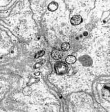
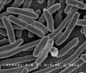
In 1924, Louis de Broglie realized that the wavelength of an electron was .05 angstroms or 1/100,000 of the wavelength of light. Eriom Schrodinger, in 1926, put the mechanical and optical components together. These discoveries helped to create the electron microscope which revolutionized looking at the microworld. Two major types of microscopes use electrons to help look at specimens in a more detailed way. In 1931 Ernst Ruska and Max Knoll created a microscope that used a beam of electrons to pass through a thin slice of a specimen. The image was focused by a magnetic coil and then onto a photographic plate, producing a higher resolution image than a compound microscope. This was the first Transmission Electron Microscope (TEM). TEM uses high voltage electron beam that is partially transmitted through a thin specimen. In 1938 the first Scanning Electron Microscope (SEM) was built by Manfred von Ardenne of Germany. He laid the foundations for SEM in several papers published in 1938. However, during the World War II his apparatus was destroyed, and he discontinued his work. Sir Charles Oatley of Cambridge, United Kingdom perfected the SEM in the 1940's. The SEM uses secondary electrons which are emitted by the surface of the specimen. The electrons are detected and mapped as an image. SEM produces a better 3D structure of a sample while the resolution of the TEM is of higher quality.
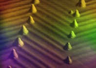
The most powerful of optical tools today are instruments that look at the atomic level of surfaces. The Scanning Tunneling Microscope (STM) provides a three-dimensional view of most metal surfaces. An electron cloud is found on metal surfaces and atoms can tunnel through the cloud for an atomic view. This technique does not use optical properties as in the previously described microscopes. It was invented by Heinrich Rohrer and Gerd Binnig in 1981 at the IBM Zurich Labs in Switzerland. Other microscopes using atoms includes Atomic Force Microscopes (AFM), Magnetic Force Microscope (MFM), Scanning Near-field Optical Microscope, and Spin Polarized Scanning Tunneling Microscope (SP-STM).
Lets go back into time and think about a world that did not understand the microscopic world. Many of the people that invented and perfected the microscope did not think about practical applications or uses of the microscope. They were more curious about microbes or little critters and describing them. The mystery that surrounded humans before the discovery of the microscope was great. How could people understand how blood flowed without looking at the details of the circulatory system? They couldn't! The microscope opened so many new avenues that today we forget the wonder of its invention. The use of the microscope historically impacted the biological sciences the most. |