BACKGROUND:
Digested food and other nutrients that are needed by
our bodies are carried to the cells by the blood. The blood also
carries wastes away from the cells. Blood must be kept moving through
the body to accomplish these jobs. This is made possible by
the heart. The heart acts as a pump. Blood is carried through
tubes called arteries, veins, and capillaries. This entire network
of blood is called the circulatory system.
The human heart contains four chambers. The upper two are
the right and left atria. The lower two are the right and left ventricles.
Arteries carry blood away from the heart and veins carry blood to the heart.
Capillaries connect arteries and veins. The heart is a mechanical
device whose function is to pump blood through the body to replenish oxygen
lacking blood cells with oxygen. The importance of constant circulation
of blood throughout the body cannot be over emphasized, for without blood,
life could not be. If blood does not reach the brain for just five
seconds an individual loses consciousness; after 15-20 seconds the body
begins to convulse; and if such a block lasts for nine minutes, irreparable
damage to the brain results.
The strong contraction of the heart muscle forces blood into
the arteries in two ways: the first expands the muscular walls of
the arteries and the second pushes the blood through the arteries to regions
of the body away from the heart. This rhythm makes the arteries expand
thereby producing a pulse. In the wrist, the pulse is detected by
a throbbing sensation near the surface.
PROCEDURE:
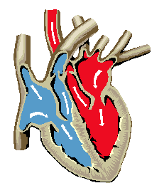
- The heart is composed of four chambers, two upper
chambers, the right and left atria, and two lower ones, the right and left
ventricles. Using the worksheet go over the heart with the students,
so that when they do the lab exercise they will be able to visualize the
heart as it creates a pulse. Point out on the smaller diagram where the
heart is located.
- Use the model of a heart for students to see
the different parts.